Australian system promises tenfold productivity increase
Laboratory scientists and clinical pathologists currently spend hours in darkened rooms analyzing and testing tissue and fluids of patients for evidence of disease. Unlike blood tests, which are performed using automated analyzers, pathology samples are subject to time-consuming manual microscopy. Digital pathology, which uses a computer-controlled microscope to scan and produce images of the samples, has helped advance what is often tedious and subjective work. But it has its problems and is not widely adopted. Now scientists in Australia are ready to roll out an AI-assisted form of digital pathology that could finally make the technique worthwhile, increasing productivity as much as tenfold.
“There has been virtually zero adoption of digital pathology outside of research and educational use,” says Brian Lovell, a professor at the School of Electrical Engineering and Computer Science at the University of Queensland, in Australia. “Low resolution and poor imaging accuracy means pathologists must manually rescan the samples many times.”
“Effectively, it predicts the areas pathologists will want to look at in high magnification, and then it enables them to zoom into those areas and see the results without having to go back to the glass slide”—BRIAN LOVELL, UNIVERSITY OF QUEENSLAND
Other experts note that the challenges extend well beyond the technical. “Widespread implementation of digital diagnostics requires significant investment in terms of personnel, financial, and IT resources, and in lab reorganization,” points out Heather Dawson, a senior staff pathologist at the Institute of Tissue Medicine and Pathology at the University of Bern, in Switzerland.
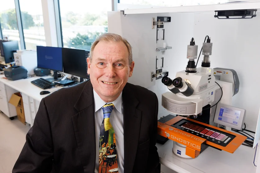
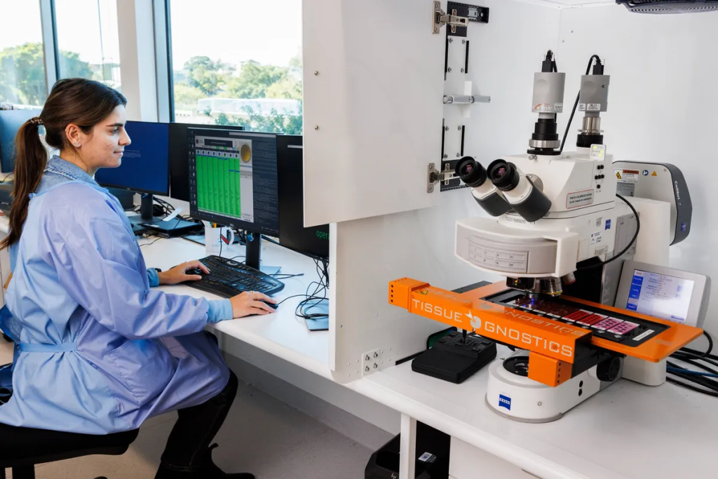
The University of Queensland’s professor Brian Lovell poses in front of the AI-based digital pathology system operating at SNP’s Brisbane lab.THE UNIVERSITY OF QUEENSLAND
But now, after 10 years of research, this could be about to change. Lovell, working with scientists at Sullivan Nicolaides Pathology (SNP), a major Australian diagnostic testing laboratory, are introducing an AI-based digital pathology system they say overcomes these hurdles. It offers as much as a tenfold increase in productivity, depending on the kinds of tests done. The technology has been accredited for commercial use by Australia’s National Association of Testing Authorities, and it has been in clinical use at SNP for several years, enabling the team to develop and perform a portfolio of 18 accredited and widely used tests.
Crucially, those accredited tests include “Gram stain testing used to identify harmful bacteria, which comprises about 50 percent of clinical microscopy tests,” says Lovell. “Without Gram, you can’t expect digital pathology to be successful commercially.”
At the center of the system are AI algorithms used to examine the images while slides are being scanned. Lovell says the system has been trained on terabytes of diagnostically classified image data, so as to seek out what a pathologist would look for in a particular kind of test—certain bacteria types, for instance. It identifies and marks areas of diagnostic interest in the image and then adjusts the microscope to rescan those areas at up to 100x magnification to create a zoomable image for the pathologist to review.
“The system doesn’t scan blindly like a photocopier,” says Lovell. “Effectively, it predicts the areas pathologists will want to look at in high magnification, and then it enables them to zoom into those areas and see the results without having to go back to the glass slide.”
But in introducing AI into pathology, Dawson raises concerns that pathologists could become overly dependent on the technology. “How much will diagnoses made by AI in a first-read setting influence pathologists’ own diagnoses?” she asks. “How well trained is the system to think outside the box; for instance, if there are unexpected findings during routine biopsies? And could pathologists miss these because they’ve become too dependent on the algorithm?”
Even without AI, digital pathology has other advantages. By using an interface like Google Earth, Lovell notes that an image can be accessed remotely anywhere in the world—to get a second opinion, for example. Archiving is another major benefit. Pathologists can now access saved images in a database at any time—legal reasons being one example—whereas the traditional method of referring back to thousands of physical glass slides, which may decompose over time, has always been a time-consuming, even impossible chore.
Lovell notes that the system’s software has been written to work with any brand of microscope, and it supports both types of microscopy used by pathologists: bright field, where the sample is illuminated by white light, and fluorescent scanning where the sample is stained with fluorescent dyes and exposed to ultraviolet light.
Along with other efficiency and procedural improvements the AI system has led to greater diagnostic accuracy, faster turnaround, and fewer sampling errors, says Lovell. “When you consider 70 percent of disease diagnoses around the world are confirmed using pathology tests, changes like these will impact the entire health care system,” he says.